
March 5, 2026
2 my read
 Add us on GoogleAdd SciAm
Scientists created a digital library full of ants
Using a synchrotron-powered CT scanner, the Antscan project created an open source digital library cataloging thousands of 3D ant specimens
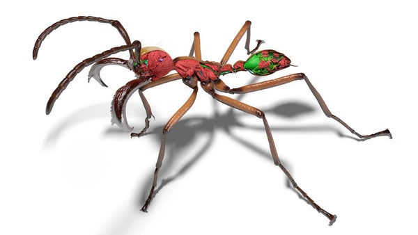
Scientists created 3D renderings of hundreds of ant species, including this one Eciton hamatum.
Ants are among the most dynamic of the “little things that rule the world,” as the late biologist EO Wilson described insects. They build complex societies, travel widely and are ubiquitous. There are more than 20 quadrillion ants out there—so many that it’s hard to get a handle on how varied the behavior and body structure of different ant species can be.
To better understand biodiversity within the ant family, researchers used a particle accelerator to create Antscan, a digital library full of three-dimensional scans and morphological data from 2,193 individual ants. The work, shared in a study published today in natural methods, sheds light on the ant’s anatomy and also on how new computer and imaging technologies can speed up research into biodiversity.
“We’re just at the beginning of even looking at the data,” says one of the study’s senior authors, Evan Economo, an entomologist at the University of Maryland, College Park. “There’s a lot of other things you can do with the project, and I’m sure there’s some really amazing stuff in there that people will dig up.”
On supporting science journalism
If you like this article, please consider supporting our award-winning journalism by subscribes. By purchasing a subscription, you help secure the future of impactful stories about the discoveries and ideas that shape our world today.
Before creating the scans, researchers collected samples of ethanol-preserved ants from museums and personal collections around the world. To capture the wide range of ant traits, they selected individuals from 212 different genera. More than 90 percent of all ant species described belong to one of the genera represented in the study.
Ant samples preserved in ethanol and scanned by the synchrotron before the researchers began to see the 3D results of their work.
Instead of the usual computed tomography (CT) scans used to image samples, researchers opted for a faster approach that would provide more detailed images by using a type of particle accelerator known as a synchrotron. Synchrotrons – such as the Karlsruhe Institute of Technology Light Source in Germany, which was used by the researchers – accelerate charged particles moving around a curved track. As the particles race around the track, they emit bright X-rays that can quickly and deeply penetrate even the smallest objects.
Each scan from the synchrotron took only seconds, but generated approximately 3,000 X-ray images of the ants.
“We were happy that we could process all the samples, but it took months before we saw the first results, and that’s when you really start to realize the magnitude of what you’ve achieved,” says Julian Katzke, one of the study’s lead authors and now a postdoctoral fellow at the Smithsonian Institution’s National Museum of Natural History.
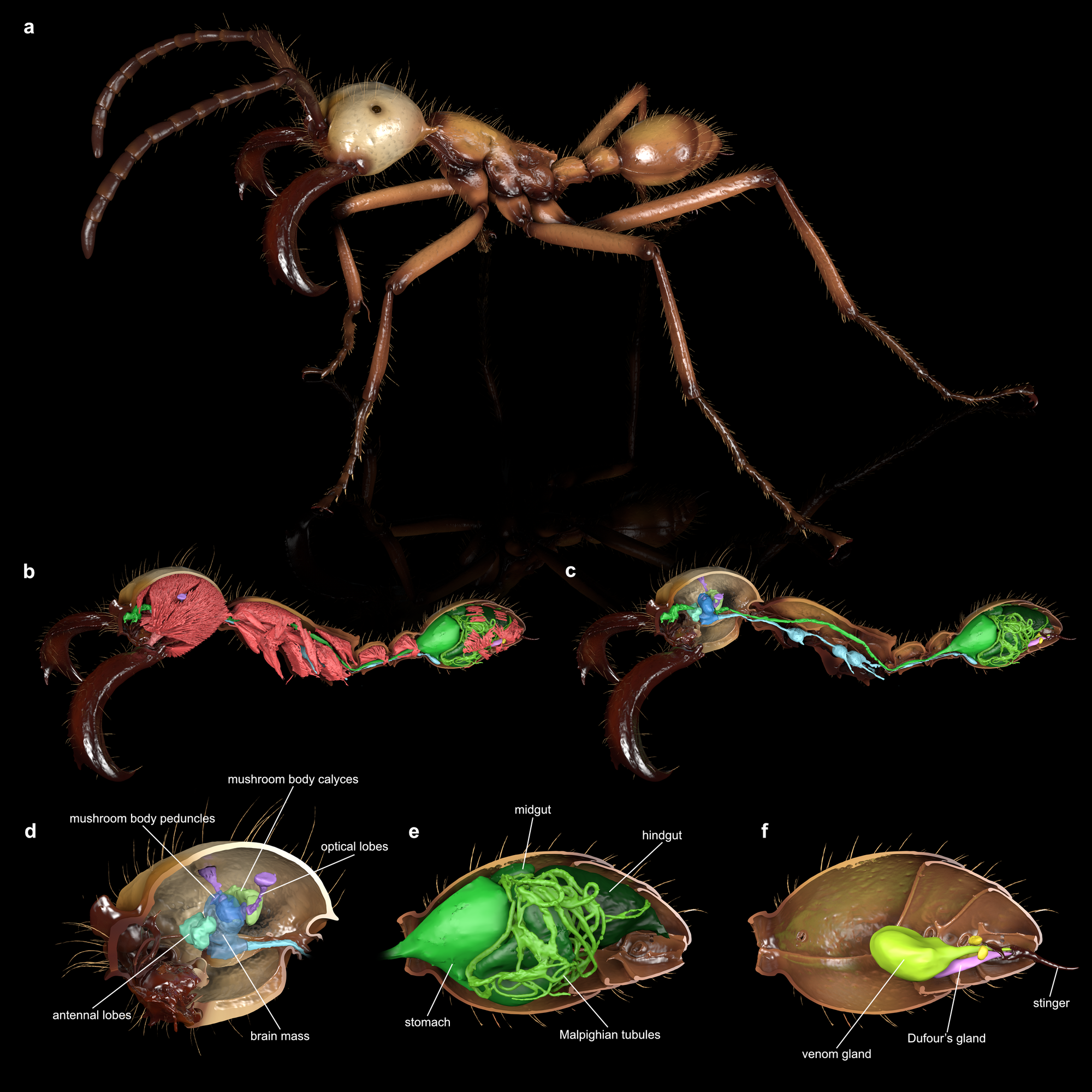
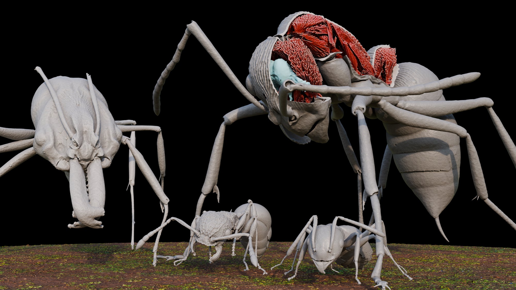
The powerful synchrotron scans provide data on the ant’s body structures – inside and out.
The result was hundreds of in-depth models assembled from layers of images showing the ants’ exoskeletons, muscles, nervous system and digestive tracts. Even parasites and strange, previously unknown anatomical features may emerge from the publicly available data when the team and other researchers fully analyze it.
And Antscan can serve as a blueprint for similar digitization projects with other insects that can reveal the similar and different characteristics between them, providing a more complete picture of insect evolution.
“This project isn’t just about, ‘Okay, we’ve got a bunch of ant scans,'” says Economo. “It points the way toward scaling this up to, eventually, all species.”
It’s time to stand up for science
If you liked this article, I would like to ask for your support. Scientific American has served as an advocate for science and industry for 180 years, and right now may be the most critical moment in its two-century history.
I have been one Scientific American subscriber since I was 12 years old, and it helped shape the way I see the world. SciAm always educates and delights me, and inspires a sense of awe for our vast, beautiful universe. I hope it does for you too.
If you subscribe to Scientific Americanyou help ensure our coverage is centered on meaningful research and discovery; that we have the resources to report on the decisions that threaten laboratories across the United States; and that we support both budding and working scientists at a time when the value of science itself is too often not recognised.
In return, you receive important news, captivating podcasts, brilliant infographics, can’t-miss newsletters, must-see videos, challenging games, and the world of science’s best writing and reporting. You can even give someone a subscription.
There has never been a more important time for us to stand up and show why science is important. I hope you will support us in that mission.


